Inadequate aldosterone produces disturbances of sodium, potassium, and water metabolism. Cortisol deficiency produces abnormal fat, protein, and carbohydrate metabolism. Absence of cortisol during a period of stress can precipitate addisonian (acute adrenal) crisis, an exaggerated state of adrenal cortical insufficiency, which is fatal if not immediately treated.
Physical findings may include the following:
- Patients with acute adrenal insufficiency generally present with acute dehydration, hypotension, hypoglycemia, or altered mental status. These signs usually occur in an acutely ill patient with sepsis or disseminated intravascular coagulation or in a patient after a traumatic delivery.
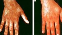
- Patients with chronic adrenal insufficiency may have increased skin pigmentation, particularly in the areolae and genitalia, as well as any scars or moles. Recent scars are typically affected most often. Areas unexposed to sun (eg, palmar creases, axillae, areolae) are often hyperpigmented, which may help to distinguish hyperpigmentation from sun tan. The patient may also have pigmentary lines in the gums. See the images below.
- Signs of weight loss may be evident. If the patient is not frankly hypotensive, he or she may have orthostatic hypotension.
- Some patients lose pubic and axillary hair because adrenal androgens support growth of body hair in these areas.
- Wolman disease (OMIM 278000), an autosomal recessive disorder caused by a deficiency of lysosomal acid lipase, may present with adrenal calcifications. Adrenal calcifications may be seen on plain radiography or CT scanning of the adrenal glands.


No comments:
Post a Comment